Guides
Melanotan I and II: structure, analytics and handling in research
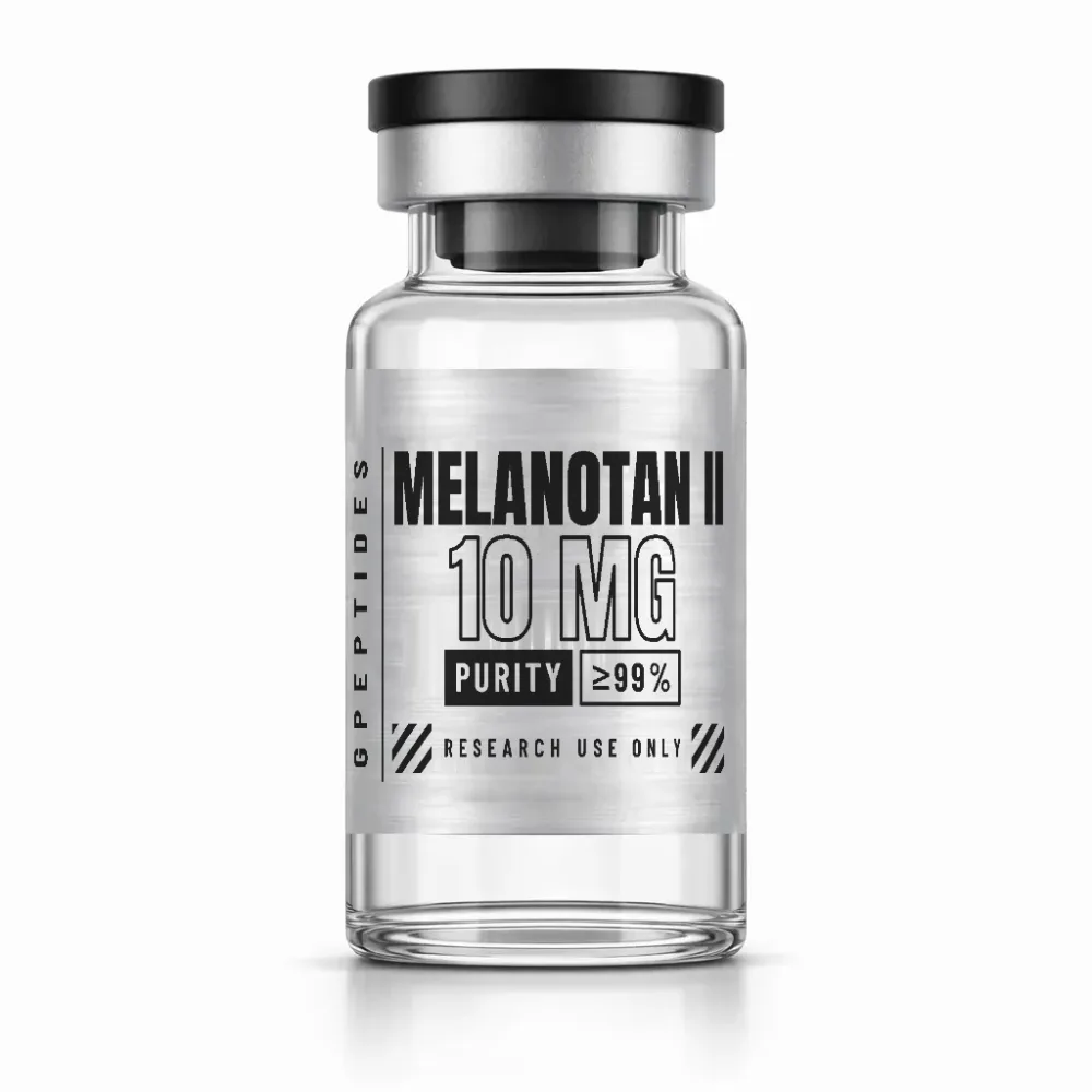
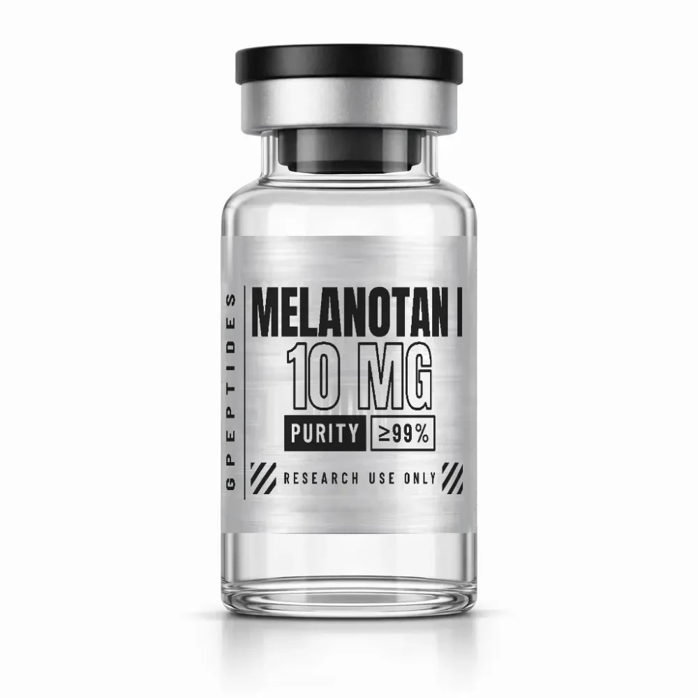
MT1 and MT2 are two structurally distinct analogues of α-MSH: a linear chain of 13 residues and a heptapeptide closed by a lactam bridge. This guide explains the naming, structure, expected masses, typical by-products and storage of the lyophilisates.
Research Use Only- Published2026-09-24
- Updated2026-09-24
- AuthorGPeptides editorial team (operator)
Overview: what lies behind MT1 and MT2
Melanotan I and Melanotan II are trivial names for two synthetic peptides, both derived from α-melanocyte-stimulating hormone (α-MSH). The catalogue lists them as MT1 and MT2; publications also use MT-I, MT-II or MTII. The Roman numerals do not denote generations of the same molecule but two separate compounds with different molecular formulae.
For MT1, the INN afamelanotide and the designation NDP-MSH are also in common use. Care is needed when searching the literature: the abbreviations MT1 and MT2 also stand for the two melatonin receptors and for isoforms of the metallothioneins. A search becomes unambiguous via the CAS number: 75921-69-6 for MT1, 121062-08-6 for MT2.
Structure in detail
The positions follow the numbering of natural α-MSH. Both peptides share three features:
- Norleucine at position 4: this non-proteinogenic amino acid is isosteric with methionine but contains no oxidisable thioether sulfur.
- D-phenylalanine at position 7: the inverted configuration lies in the middle of the tetrapeptide His-Phe-Arg-Trp, which the literature regards as the central recognition motif of the melanocortin receptors.
- Protected termini: the N-terminus is acetylated and the C-terminus amidated. This removes the free amino group and the free carboxyl group at which amino- and carboxypeptidases act.
MT2 goes two steps further. It comprises only positions 4 to 10, and positions 5 and 10 are occupied by aspartic acid and lysine. Their side chains are joined with loss of water to form a lactam; the macrocycle has 23 ring atoms, recognisable in the IUPAC name from the hexazacyclotricosane skeleton. The ring holds the recognition motif in a few preferred conformations. Because both termini are also blocked, the only ionisable side chains left in MT2 are histidine and arginine; MT1 additionally carries a free lysine, a glutamic acid and a tyrosine.
Distinction from related molecules
- α-MSH: the natural model, linear, 13 residues, with methionine at position 4 and L-phenylalanine at position 7.
- MT1: linear, 13 residues, C78H111N21O19, around 1646.8 g/mol; linked in the literature above all with the MC1 receptor.
- MT2: cyclic, 7 residues, C50H69N15O9, around 1024.2 g/mol; described as a non-selective agonist of several melanocortin receptor subtypes.
- SHU9119: not a catalogue product, but a frequently cited reference molecule: MT2 with D-2-naphthylalanine at position 7 (C54H71N15O9), described as an antagonist at the MC3 and MC4 receptors. Here a single residue shifts the receptor profile.
- KPV: the unmodified tripeptide of positions 11 to 13 as the free acid; contained as a sequence section in MT1 but not in MT2.
- GHK-Cu: the same catalogue category, but a copper–peptide complex with no connection to α-MSH.
What to look for in analytics and the COA
In electrospray mass spectrometry, several charge states appear, depending on the number of protonatable groups (monoisotopic, rounded):
- MT2 (M = 1023.54 Da): [M+H]⁺ at m/z 1024.55, [M+2H]²⁺ at m/z 512.78, sodium adduct [M+Na]⁺ at m/z 1046.53.
- MT1 (M = 1645.84 Da): [M+2H]²⁺ at m/z 823.93 and [M+3H]³⁺ at m/z 549.62; depending on the measurement conditions, [M+H]⁺ at m/z 1646.84 also occurs.
By-products give themselves away through their mass difference:
- Deletion sequences: lighter by the mass of one residue, for example minus 186 Da without tryptophan.
- Missing acetylation: minus 42 Da.
- Free acid instead of amide: plus just under 1 Da; easy to overlook without high-resolution measurement.
- Oxidation: plus 4, 16 or 32 Da. Because norleucine replaces methionine, the tryptophan residue is the most likely point of attack in both molecules.
- MT2 only: the unclosed precursor (plus 18 Da) and cyclic dimers. The doubly charged dimer lies at the same m/z as the singly charged monomer; it can be distinguished by the halved isotope spacing or by its retention time.
- Diastereomers: for example with L- instead of D-phenylalanine at position 7. As the mass is identical, only chromatography separates them.
HPLC purity states the area share of the main peak, not the peptide share of the fill weight. Synthetic peptides are present as salts, usually as acetate or trifluoroacetate (TFA); counter-ions and residual water add to the mass in the vial without entering into the area percentages. The counter-ions are bound by arginine and histidine in MT2, and additionally by the lysine in MT1. This share is captured by the net peptide content, a quantity independent of purity. MT1 and MT2 are regularly tested externally by HPLC and mass spectrometry. The available certificates of analysis (COA) are published in the lab area; as long as no certificate has been filed there, testing is still pending. For more depth, see the guides HPLC and mass spectrometry, Reading a certificate of analysis and Purity of research peptides.
Storage and handling in the laboratory
As lyophilisates stored dry, dark and cool, both peptides are considerably more stable than in solution; for longer periods, the freezer is the natural choice. Points to note:
- Light and oxygen: tryptophan is light-sensitive and prone to oxidation. Keep the vials protected from light and open them only briefly.
- Moisture: lyophilised peptide salts are hygroscopic. Let a chilled vial come to room temperature while still sealed before opening it, so that no condensation settles on the powder.
- Solutions: these are less stable than the lyophilisate; you should avoid repeated freezing and thawing.
The ring makes MT2 more rigid but protects it neither from oxidation nor from moisture. General notes are given in the guide Storing lyophilised peptides.
Research context
Both molecules are studied in in-vitro and preclinical research on the melanocortin system. MT1 is used above all in cell models to study the signalling cascade of the MC1 receptor; radiolabelled NDP-MSH is also a widely used tracer in binding assays. Because of its broad profile, MT2 serves as a reference agonist for comparing signals at the MC1, MC3, MC4 and MC5 receptors, often together with antagonists such as SHU9119.
Research use only: legal notice
At GPeptides, MT1 and MT2 are placed on the market exclusively as laboratory reagents for in-vitro research. They are not intended for use in or on the human or animal body and are offered neither as medicinal products nor as cosmetic products. GPeptides makes no statements on use or effects. Fill weight and price are given on the product pages MT1 and MT2; further related molecules are listed in the Melanocortins & Copper Peptides category.
Related categories: Melanocortins & Copper Peptides
Related products
Related guides
HPLC and mass spectrometry: basics of peptide analytics
Two methods, two questions: HPLC answers “how pure?”, mass spectrometry “is it the right molecule?”. What lies behind peak, retention time and m/z.
Read more: HPLC and mass spectrometry: basics of peptide analytics →How to read a certificate of analysis (COA)
Test method, purity in percent, test date, laboratory: a COA is only as good as the information it contains. How to check whether a certificate matches the vial in your hand.
Read more: How to read a certificate of analysis (COA) →Purity of research peptides: what the percentages mean
Purity is not the same as content. Anyone comparing COAs should be able to distinguish area percent, net peptide content and typical by-products of peptide synthesis.
Read more: Purity of research peptides: what the percentages mean →