Guides
Cagrilintide: structure, analytics and handling in research
Cagrilintide is a lipidated analogue of human amylin comprising 37 amino acids, with six substitutions, a disulfide bridge and a C20 fatty diacid bound via γ-glutamic acid. This guide explains its naming, its structure and how it differs from amylin, pramlintide and calcitonin, as well as the analytics and storage of the laboratory reagent.
Research Use Only- Published2026-09-24
- Updated2026-09-24
- AuthorGPeptides editorial team (operator)
Overview and naming
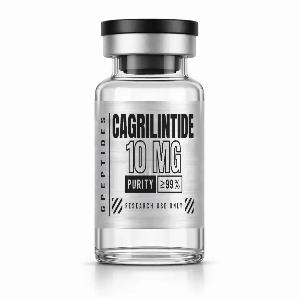
Cagrilintide (spelt Cagrilintid in German), which is also the name used in the catalogue, is the international non-proprietary name (INN) of a synthetic peptide derived from human amylin. The WHO published the name in 2020 in List 123 of proposed INNs, with the class designation amylin analogue. GPeptides supplies the reagent as a lyophilisate in a vial containing 10 mg.
- CAS number: 1415456-99-3
- UNII: AO43BIF1U8
- PubChem CID: 171397054
- Molecular formula: C194H312N54O59S2, molar mass around 4409.1 g/mol
Take care when comparing databases: the computed IUPAC name at PubChem gives D-configurations for several residues between positions 27 and 37, whereas the INN, the CAS name and the GSRS substance register describe L-amino acids throughout. You should therefore not use the SMILES and InChIKey from this entry without verifying them.
Structure in detail
As in human amylin, the backbone comprises 37 amino acids, written in blocks of ten: KCNTATCATQ RLAEFLRHSS NNFGPILPPT NVGSNTP-NH2. Four features characterise the molecule:
- Six substitutions relative to human amylin: N14E, V17R, A25P, S28P, S29P and Y37P. Glutamic acid 14 and arginine 17 are three positions apart on the same face of the helix; their oppositely charged side chains are described as a helix-stabilising salt bridge. Human amylin tends to form amyloid fibrils, hence its second name IAPP (islet amyloid polypeptide); the prolines at 25, 28 and 29, adopted from pramlintide, fit poorly into β-sheets.
- Disulfide bridge: Cys2 and Cys7 close residues 2 to 7 into a 20-membered ring. The WHO numbers the residues according to the precursor protein and writes ‘(35-40)-disulfide’; this refers to the same bridge.
- Lipidation: attached to the α-amino group of lysine 1 via an L-γ-glutamyl linker is the monoamide of eicosanedioic acid, an unbranched C20 dicarboxylic acid. The hydrocarbon chain makes the molecule amphiphilic; the carboxyl group at the end of the chain, the α-carboxyl group of the linker and the ε-amino group of the lysine remain ionisable.
- C-terminus: prolinamide instead of tyrosinamide; the chain therefore no longer contains any tyrosine.
By calculation, at neutral pH lysine 1, arginines 11 and 17 and, in part, histidine 18 are offset by three carboxylates: glutamic acid 14, the linker and the end of the fatty acid. The net charge is close to zero.
How it differs from related molecules
- Human amylin (IAPP, INN amlintide): 37 residues, Cys2–Cys7, tyrosinamide at the C-terminus, no lipidation.
- Pramlintide: human amylin with A25P, S28P and S29P. Cagrilintide adopts these prolines and adds N14E, V17R, Y37P and the fatty diacid.
- Salmon calcitonin: 32 residues, disulfide bridge Cys1–Cys7, likewise prolinamide at the C-terminus; a common reference ligand in receptor-binding studies.
- Incretin analogues such as GLP-3: these address GLP-1, GIP and in some cases glucagon receptors. GLP-3 shares the C20 fatty diacid with cagrilintide, but not its sequence or target receptor.
What to look for in analytics and the COA
- Expected mass: 4406.25 Da monoisotopic, around 4409.1 Da average. By calculation, at just over 4.4 kDa the M+2 peak is around three times as intense as the monoisotopic one; you should therefore check what a deconvoluted value refers to. A database entry with O58 is a typographical error; only O59 matches the mass given there.
- Charge states: without a free N-terminus, low charge states are plausible with electrospray ionisation; based on the average mass, [M+3H]3+ at m/z ≈ 1470.7 and [M+4H]4+ at m/z ≈ 1103.3.
- Disulfide bridge: the reduced, open form is 2.02 Da heavier, which in the quadruply charged ion amounts to only 0.5 m/z. With a single bridge, intramolecular rearrangement is ruled out, but dimers can form via disulfide exchange. Their octuply charged ion falls at the same m/z as the quadruply charged monomer, but shows isotope peaks spaced 0.125 instead of 0.25 m/z apart and can also be distinguished by its retention time.
- Lipidation: without the fatty diacid, the mass is 324.27 Da lower; without the fatty diacid and linker, 453.31 Da lower. The acyl chain markedly prolongs retention in reversed-phase HPLC.
- Deamidation and deletion sequences: five asparagines and glutamine 10 can deamidate (+0.98 Da); the particularly susceptible Asn-Gly motif is absent. Deletion sequences from the synthesis are, for example, 97.05 Da (proline) or 114.04 Da (asparagine) lighter.
- Oxidation and UV: methionine, tryptophan and tyrosine are absent. A signal at +16 Da is therefore rarer than with many peptides; the molecule barely absorbs at 280 nm, so purity is usually determined at 210 to 220 nm.
- Counter-ions: trifluoroacetate (TFA) or acetate bind to lysine, the arginines and, where present, histidine; four bound acid molecules increase the formula mass by around 456 g/mol (TFA) or 240 g/mol (acetic acid) respectively. HPLC purity as the area share of the main peak does not capture this; net peptide content refers to the total weighed quantity.
The identity and purity of Cagrilintide are regularly tested externally by HPLC and mass spectrometry. The available certificates of analysis (COAs) can be found in the lab area; where no certificate has been filed, the test is marked there as ‘pending’. How to interpret the chromatogram and mass spectrum of a certificate is explained in the guides HPLC and mass spectrometry and How to read a certificate of analysis.
Storage and handling in the laboratory
Store the lyophilisate dry, cool and protected from light, and at −20 °C or colder for longer periods. Let a refrigerated vial reach room temperature while still closed before opening it, so that no condensation forms. Specific to this molecule:
- Amphiphilicity: lipidated peptides may tend to self-associate in solution and adhere to surfaces.
- Reducing agents: DTT or TCEP open the disulfide bridge and are unsuitable for experiments with the intact molecule.
- pH: alkaline conditions promote disulfide exchange and deamidation.
It is better to aliquot solutions than to freeze and thaw them repeatedly. More on this in the guide Storing lyophilised peptides.
Research context
The research field of cagrilintide is the amylin and calcitonin receptor system, studied in vitro and in preclinical models. The calcitonin receptor (CTR) is a class B G-protein-coupled receptor; together with RAMP1, RAMP2 or RAMP3 it forms the amylin receptors AMY1R, AMY2R and AMY3R. Cagrilintide is described as a non-selective agonist at CTR and all three amylin receptors; topics studied include binding and signal transduction in cell systems.
Research use only: legal notice
At GPeptides, Cagrilintide is available solely as a laboratory reagent for in-vitro experiments. It is not intended for use in humans or animals, and it is not sold as a medicinal product. GPeptides makes no statements on use or effects; the test reports document identity and purity alone. Price and availability are shown on the product page Cagrilintide, and related molecules in the Incretin Analogues category.
Related categories: Incretin Analogues
Related products
Related guides
HPLC and mass spectrometry: basics of peptide analytics
Two methods, two questions: HPLC answers “how pure?”, mass spectrometry “is it the right molecule?”. What lies behind peak, retention time and m/z.
Read more: HPLC and mass spectrometry: basics of peptide analytics →How to read a certificate of analysis (COA)
Test method, purity in percent, test date, laboratory: a COA is only as good as the information it contains. How to check whether a certificate matches the vial in your hand.
Read more: How to read a certificate of analysis (COA) →Storing lyophilised peptides: temperature, light, humidity
Dry, cold, dark: lyophilisates are robust but not indestructible. The key rules for unopened vials, opened vials and solutions.
Read more: Storing lyophilised peptides: temperature, light, humidity →